Posts

Arthritis of the Knee: An Overview

Image
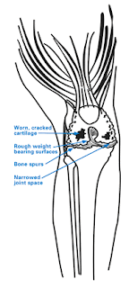
Introduction: Anatomy of the Knee The knee joint is a structure composed of three bones: the femur (thigh bone), the tibia (shin bone) and the patella (knee cap). The bones are covered with smooth cartilage surfaces that act as a cushion during weight-bearing activity. The bones of the knee are connected by strong ligaments and powerful muscles that are attached to the thigh and calf by tendons and provide side-to-side stability. In a healthy knee, all of these structures work together to allow the knee to flex (bend) and extend (straighten) the lower leg smoothly. Arthritis is a disease that affects the surface of the joint (cartilage) wearing down so it no longer moves smoothly and loses the ability to acts like a cushion. Damaged cartilage causes a roughened surface and may lead to bones rubbing directly together, causing persistent pain, clicking, a catching sensation, and limited range of motion. Knee Arthritis There are three common types of arthritis: Oste...

Shoulder Osteoarthritis Symptoms

Image
The symptoms of shoulder arthritis tend to progress gradually; sometimes shoulder osteoarthritis symptoms may seemingly get better, only to return. Many people initially attribute shoulder soreness or stiffness to lack of exercise or getting older. However, shoulder arthritis can progress and eventually interrupt one’s ability to enjoy everyday activities. Early recognition of symptoms and appropriate activity modification and treatment can sometimes slow or eliminate progression of shoulder osteoarthritis symptoms. Joint tenderness and pain. Tenderness and pain due to glenohumeral osteoarthritis is usually centered at the back of the shoulder and can be felt in the middle-range of motion rather than at the extremes of flexibility. Those with moderate to severe glenohumeral arthritis may experience dull aching pain at night making it difficult to sleep. Pain may intensify with changes in the weather. Stiffness or loss of motion. Aside from pain, a major symptom of sh...

Preoperative orientation

Image
Most patients will be asked to donate 1 to 2 pints of their own blood in the weeks preceding knee replacement surgery. This helps reduce the need for a blood transfusion from our blood bank. Almost all of the patients will receive the donated blood as a transfusion after surgery. Rarely, an additional transfusion is necessary from our blood bank. The blood from the blood bank is carefully screened to the best of our ability to detect any infectious diseases. You will be asked to see a medical doctor at HSS prior to your surgery. This is a precaution to make certain that you are healthy enough to undergo knee replacement surgery. In the course of this workup, you may be asked to have additional testing to examine your heart and lung function. After your surgery, this medical doctor will see you in the hospital. Postoperative course Immediately after total knee replacement surgery, you will be in the recovery room. Most patients are able to go to a regular room after a fe...

Total knee replacement

Image
History   Total knee replacement surgery was first performed in 1968, and has evolved over the years into a reliable and effective way to relieve disabling pain and allow patients to resume their active lives. Improvements in surgical techniques and implant design and construction have helped make this one of the most successful orthopaedic procedures today. As the population has become older and remained more active, the need for total knee replacement continues to increase. Today, approximately 270,000 total knee replacements are done every year in the United States. Many of the advancements in knee replacement surgery have taken place at HSS. Improvements in the surgical technique and the design of new implants are a few of the contributions the surgeons of the Hip and Knee service have made. Decision to have surgery People often wonder when and why they should have their knee replaced. This is an individualized question that depends upon a person's activit...

Treatment of arthritis of the knee

Image
Depending upon the severity of arthritis and the patient's age, knee arthritis may be managed in a number of different ways. Treatment may consist of operative or non-operative methods, or a combination of both. Non-operative The first line of treatment of knee arthritis includes activity modification, anti-inflammatory medication, and weight loss. Giving up activities that make the pain worse may make this condition bearable for some people. Anti-inflammatory medications such as ibuprofen, naprosyn and newer Cox-2 inhibitors help alleviate the inflammation that may be contributing to the pain. Physical therapy to strengthen the muscles around the knee may help absorb some of the shock imparted to the joint. This is particularly true for knee-cap (patello-femoral) arthritis. Special kinds of braces, designed to place transfer load to a part of the knee that is less arthritic may also help relieve the pain. Injections of medication inside the knee joint may also help all...

Knee joint

Image
He  knee joint can be thought of as a hinge joint with the primary motion of straightening and bending. In reality, it is more complex than a simple hinge, as the surfaces actually glide and roll upon one another. It is composed of the end of the thigh bone (femur), the top of the leg bone (tibia), and the kneecap (patella). The ends of the bone are covered with a smooth, glistening layer called articular cartilage. The articular cartilage is what allows the bones to glide smoothly with less resistance than ice sliding on ice. The articular cartilage can be seen on X-ray as the space in between the bones. The knee can be thought of as having 3 compartments - the medial, the lateral, and the patellofemoral. In addition, there are 2 special cartilages within the knee joint called the lateral and medial meniscus, which act as shock absorbers within the knee joint. There are also 2 ligaments within the knee, called the anterior cruciate ligament and the posterior cru...

Types of Shoulder replacement surgery

Image
When the Non-surgical methods fail to treat Shoulder arthritis, your doctor will suggest you to opt for Shoulder joint replacement surgery. Shoulder replacement surgery is done to remove all the damaged parts and is replaced with artificially made prosthetic parts made of metals or plastics. Shoulder replacement surgery includes relieve from severe pain of arthritis and increases your ease and range of motion.  Different types of Shoulder Replacement surgeries are Total/Traditional shoulder replacement It is the most dependable option for severely damaged shoulder, it is the complete replacement of the shoulder (both ball and socket parts). After the surgery, range of motion is restored and arthritis pain is relieved. This is contraindicated if there is damage to the rotator cuff or rotator cuff tear arthropathy. Reverse Total Shoulder Replacement This is also the total replacement of ball and socket from the joint with the prosthesis but th...